As time passes, we age, and our bodies experience numerous changes. One of these changes could be how we hear things. Some people begin losing their ability to listen and require assistance from hearing aids to regain their hearing ability. This blog post will discuss sensorineural hearing loss and how it affects people. We’ll also be looking at how audiologists and otolaryngologists look into and care for underlying conditions that could lead a patient to lose hearing ability.
What Sensorineural Hearing Loss Means:
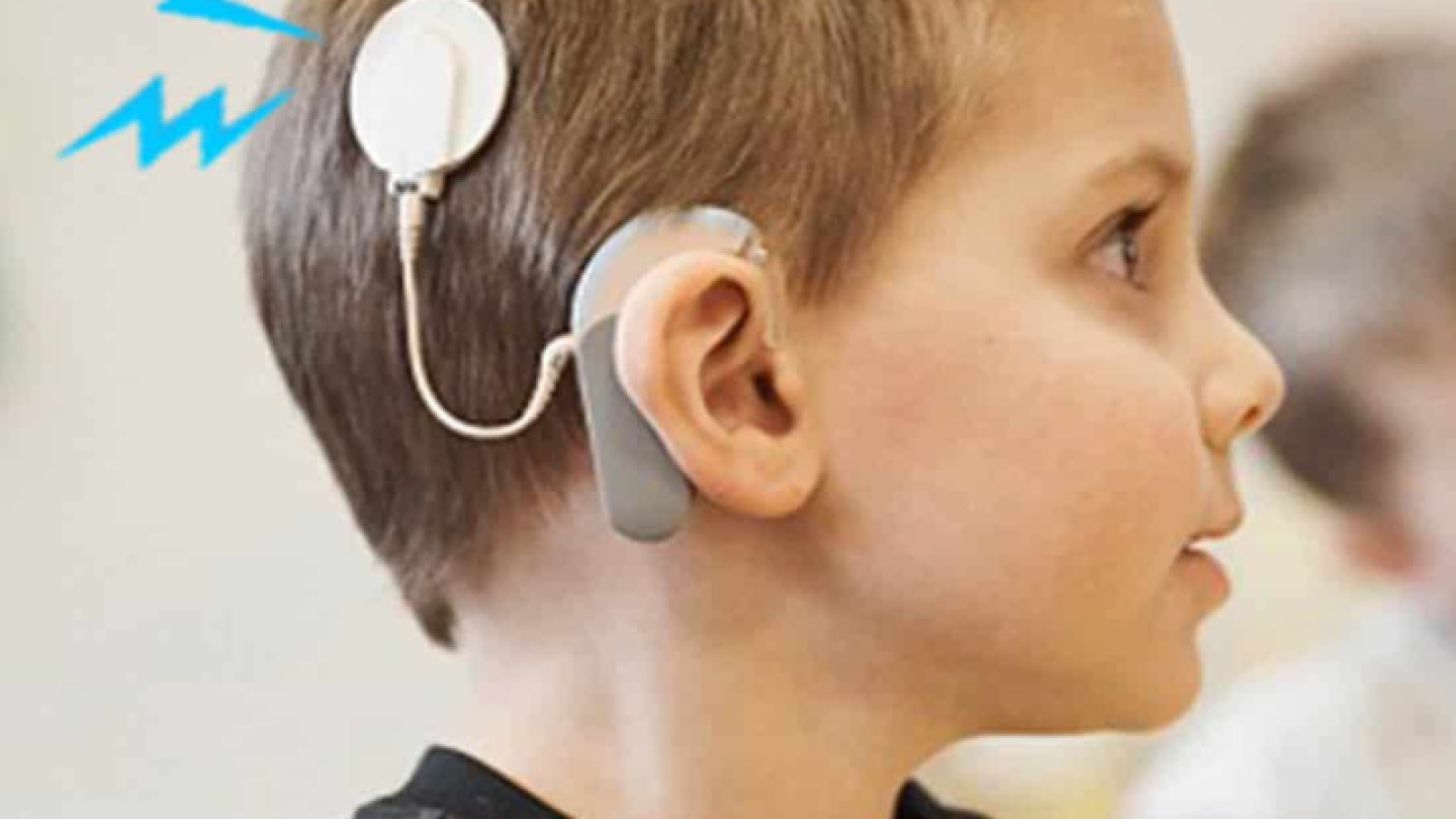
Hearing loss is one of the most frequent complaints, and referrals are commonly created for secondary care for an otolaryngologist’s attention. Only two kinds of hearing loss exist: sensorineural and conductive hearing loss. Sensorineural hearing loss (SNHL) is the most frequent hearing loss, and most of those who have lost the ability to hear are affected by this hearing loss. SNHL means any reason for hearing loss because of a pathology of the auditory nerve or cochlea and experiencing full audiometric assessment by a multidisciplinary team involving a radiologist, audiologist, otolaryngologist, and speech/language therapist.
Etiology:
The most prevalent reasons as to why sensorineural hearing loss occurs are:
Presbycusis.
Congenital – nonsyndromic and syndromic
Noise-inflicted hearing loss
Ototoxicity – aminoglycosides, a few chemotherapeutic agents, and loop diuretics.
Systemic criteria – diabetes, meningitis.
Meniere’s disease.
Vestibular schwannoma.
Other causes are autoimmune, perilymphatic fistula, and barotrauma.
Pathophysiology:
Sensorineural hearing loss often occurs from trauma caused by the hair cells found within the inner ear, the vestibulocochlear nerve, or the brain’s central processing centers. This is distinct from conductive hearing loss, as hearing loss occurs from the inability of sound waves to get to the inner ear. The ear is composed of three components: The Inner ear, the Middle Ear & the Inner ear. Every part of the ear is paramount for the conduction of sound waves, but within the SNHL, we’ll be looking at the pathology in the inner ear, which takes the sound waves to the hearing loss. The interface between the oval and stapes window delivers sound transmission of the cochlea. Once the sound has reached the cochlea, it’ll undergo the first amplification by the outer hair cells, and then electrochemical transduction occurs by the inner hair cells. The cochlea will acquire an acoustic signal, and a traveling wave shall be made, which shall travel through the cochlea’s basilar membrane that stimulates the outer hair cells (OHCs), which serve as a biological compressor/amplifier and change the signal. The basilar membrane of the cochlea shall be very frequency-specific and tonotopically organized. The base of the basilar membrane shall respond to the higher frequency noises, while the apex shall respond to the lower frequencies. The inner hair cells (IHCS) found within the cochlea shall transduce the energy of the traveling wave to an electric action potential and synapse at the spiral ganglion to create the auditory nerve. There are a lot of pathophysiological mechanisms, and damage to them could result in the inner experiencing SNHL.
Aberrant Metabolic Activity:
The transportation of ions establishes cochlear function. Any conditions acquired due to genetics or trauma could interfere with this transport, leading to transformations in the endolymph and influencing how one person hears.
Vascular:
If there is any interference with the vascular supply of the cochlea, it may result in conditions such as ototoxicity, systemic vascular situations, and noise trauma, which will affect the function of the stria vascularis.
Structural Abnormality of the Cochlear Parts:
These abnormalities might include congenital or trauma conditions.
The Basilar Membrane Being Overcrowded:
Preventing the motility and transduction of the OHCs & IHCs transduction capabilities could result in common conditions such as autoimmune pathology and diabetes.
Noise Trauma:
With noise trauma, the vibrational shift between the basilar and tectorial membrane grows, and the shift has the potential to damage the stereocilia of the OHCs. Because of this, the stiffness of the organ of Corti decreases. Aminoglycoside antibiotics, such as gentamicin, which are potassium channel blockers, can stop the hair cells from depolarizing.
Histopathology:
One of the organs of the Corti is the cochlea’s sensory epithelium. It’ll be found in the cochlea’s Scala media and comprises sensory hair cells and non-sensory supporting cells. The sensory cells are the OHCs, and the IHCS put together uniquely. One row of the IHCs and three rows of the OHCs will be separated by supporting cells. The hair bundle is made up of stereocilia rows, which grow in height within one direction across the apical surface of the hair cell. Movements occur between the basilar and tectorial membrane, which causes deflection of the stereocilia, which activates or deactivates receptors on the surface of the hair cells. Potassium ions shall go into the hair cell, which shall depolarize it, making the calcium channels open, and the auditory nerve then sends the sound wave’s information to the brain.
Conclusion:
People who have sensorineural hearing loss will have a significant part of their life affected by it. Many people are affected by this condition at some point in their lives. The best thing to do would be to acquire the care of an interprofessional team that involves primary doctors, audiologists, neuroradiologists, and otolaryngologists. These people will be able to provide the sensorineural hearing loss treatment. It’ll be essential to diagnose these symptoms early on so that there will be no long-term effects later on in the patient’s development.
