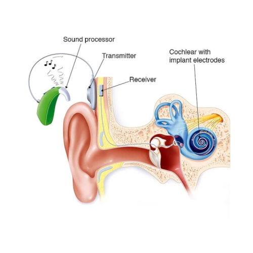
A compact electronic medical device known as a cochlear implant is intended to furnish individuals who are profoundly deaf or severely hard of hearing with a perception of sound. In contrast to hearing aides, which amplify sound, cochlear implants directly stimulate the auditory nerve and bypass impaired areas of the ear. The components and functionality of a cochlear implant are summarized below:

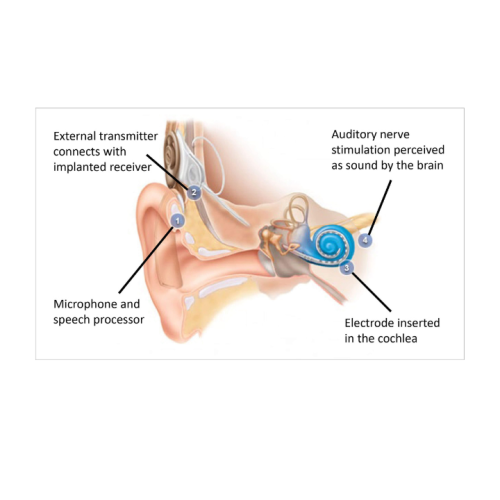
The placement of a cochlear implant, a compact electronic device that provides a sensation of sound to individuals who are profoundly deaf or severely hard of hearing, is a procedure known as cochlear implant surgery. An synopsis of the surgery is provided below:

Assessment of Medical Suitability: A comprehensive evaluation conducted by an otolaryngologist (ENT specialist) and an audiologist to ascertain eligibility.

The anatomy of the inner ear and cochlea is evaluated through imaging studies, such as MRI or CT scans.

Audiological Testing: Audiological evaluations that verify the extent of hearing loss.

Patient counseling: Providing the patient with information regarding the implant’s risks, benefits, and realistic expectations.

Surgery Preparation:

Anaesthesia: The patient is administered general anesthesia.

Patient Positioning: The patient is positioned to ensure that the surgeon has the best possible access to the ear.

 
cochlear-1
cochlear-2

Steps of Procedure

The following are the steps of the procedure

  • A tiny incision is made behind the ear.
  • The surgeon penetrates into the mastoid bone, which is a portion of the cranium located behind the ear, to gain access to the middle ear during a mastoidectomy.
  • A small opening is made in the cochlea (cochleostomy) or the round window membrane is used to insert the electrode array (round window approach).
  • Insertion of Electrode Array: The cochlear implant’s electrode array is delicately inserted into the cochlea.

Location of Receiver:

  • The internal receiver/stimulator is secured to the cranium and inserted beneath the skin.
  • Intraoperative testing is conducted to guarantee that the device is functioning properly.
  • Suture closure: The incision is closed with sutures.
  • Recovery Postoperative Care The patient is monitored in the recovery chamber until they are completely awake from anesthesia.

Pain Management:

  • Medication is normally employed to alleviate pain.
  • Initial Follow-Up: A follow-up appointment is scheduled to evaluate the surgical site and general health.
  • Activation: In a few weeks, the external components of the cochlear implant are installed, and the device is activated and programmed after the surgical site has recovered.
  • The speech processor is initially programmed and fine-tuned by an audiologist during rehabilitation.
cochlear-3
cochlear-4

Auditory Training:

The patient is exposed to auditory rehabilitation in order to enhance their comprehension of speech and to learn how to interpret new sounds.

Scheduled Follow-Up: Continuous evaluations and modifications to enhance the functionality of the cochlear implant.

Complications and Risks

Infection: There is a risk of infection at the surgical site.
Failure of the device: The potential for device malfunction or rejection.
Damage to Nerves:

  • Potential damage to facial nerves.
  • Balance issues or vertigo, which may occur postoperatively.
  • Possible alterations in taste sensation as a result of nerve proximity Taste Disturbance.
  • Predicted Results

Hearing enhancement:

  • A substantial enhancement in the capacity to comprehend speech and perceive sound.
  • An adjustment period is necessary for the brain to adjust to the new method of hearing, necessitating perseverance and practice.
  • A highly specialized procedure known as cochlear implant surgery provides numerous individuals with the opportunity to participate more actively in their daily lives and hear more clearly.
cohlear-2